The number of young people living with debilitating and incurable bowel conditions worldwide is growing, but researchers now hope a one-of-a-kind microscope will help them understand such diseases and find new treatments.
According to the British Society of Gastroenterology, more than half a million people in the UK are living with inflammatory bowel disease (IBD), with Crohn’s disease and ulcerative colitis the two main forms.
While such conditions can develop at any age, experts say they are most often diagnosed between the ages of 15 and 40, blighting lives at a crucial time for education, work and relationships.
Now a new piece of kit could bring fresh hope to those affected by IBD.
“What this microscope technology and what the science we do in the lab can bring is really to understand the mechanism [of IBD] better,” said Dr Karina Pombo-Garcia, a group leader at the Rosalind Franklin Institute at the Harwell Science and Innovation Campus, Oxfordshire, where the microscope is housed.
“With IBD, and with other many diseases that we collaborate with other groups to study, there [are] some treatments, but maybe those treatments still are not maybe the best or the most targeted that we can do because we … fundamentally don’t understand the disease fully.”
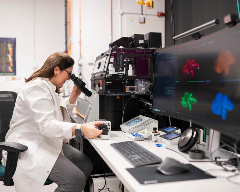
With its black casing, grey hoses and a plethora of wires, the microscope is remarkably unobtrusive.
Yet this is no run-of-the-mill bit of tech: the microscope operates at 10 times the resolution of conventional light microscopy, allowing researchers to see the details down to just 20nm in size.
To do so, the microscope, which researchers have nicknamed “Curie”, uses a two-laser technique known as stimulated emission depletion (STED) microscopy that scooped its inventors the Nobel prize in physics in 2014.
In this setup, features of interest within cells are labelled with fluorescent tags – these absorb energy from the first laser beam and then emit light, or fluorescence.
A second, doughnut-shaped laser beam overlaps with the area illuminated by the first. Crucially, this second beam “switches off” fluorescence within its outer ring, meaning fluorescence is only detected from the centre of the doughnut – a very small region.
“This allows the microscope to distinguish structures that are much closer together than would be possible using a conventional fluorescence microscope, producing images with exceptionally high resolution,” said Pombo-Garcia.
The microscope also has additional features, making it the only one of its kind in the UK. These, Pombo-Garcia notes, include deformable mirrors that can correct for optical distortions caused by the sample.
“This allows researchers to obtain clear images from deeper within relatively thick tissues and mini-organs,” she said. “It also has a temperature-controlled stage, enabling living cells and mini-organs to be studied under conditions close to normal body temperature.”
Related: Bowel disease breakthrough as researchers make ‘holy grail’ discovery
Dimitrios Ioannidis, a PhD student, is using the microscope to understand how cells that line our internal organs, including the intestines, come together and create protective barriers.
“For this to happen, we have specific protein complexes, which we hypothesise might change over time. So using this piece of equipment, we are able to visualise these complexes beyond what [we can see with a] conventional microscope,” he said, adding that he was looking at how the structure of these protein complexes changes from foetal development to adulthood. “If the structure changes, so does the function,” he said.
Ioannidis noted that by understanding what healthy tissue should look like, and then comparing and understanding the differences seen in people with disease, there was the potential to shed light on what had gone wrong.
“And then if you work and dig a little bit deeper, you can potentially find the root cause of some of the diseases that are affecting individuals,” he said, noting that this included IBD.
The microscope was unveiled by science minister Chris McDonald, alongside the announcement of £67m of funding for the Rosalind Franklin Institute over the next five years from April 2027, and a further £90m over the same period for the Henry Royce Institute, which focuses on materials research. In both cases, the funding has come from the Engineering and Physical Sciences Research Council via the core budget of UK Research and Innovation (UKRI) set in the 2025 spending review.
The announcement comes at a difficult time for UK science facilities. Earlier this year the Science and Technology Facilities Council (STFC), also part of UKRI, revealed it needs to make £162m in savings by 2030 as a result of factors such as soaring energy costs at its national facilities, increasing staffing costs, an expanded portfolio of activities and “unfavourable movements” in foreign exchange rates.
Related: World-leading UK science facilities at risk amid £162m funding crisis
The situation has put some national facilities at risk of closure, with the Diamond Light Source, Central Laser Facility and ISIS Neutron and Muon Source – all of which sit on the same campus as the Rosalind Franklin Institute – jointly facing a 15% drop in funding, leading to concerns from the scientific community.
However, McDonald maintained such facilities still receive a very high level of funding, and stressed not every project or initiative backed by the STFC will be funded in perpetuity.
“I’m not particularly happy about the characterisation of cuts because that’s not the case at the high-level budget,” he said. “But it is right that STFC live within their means and prioritise their own budget,” he said.